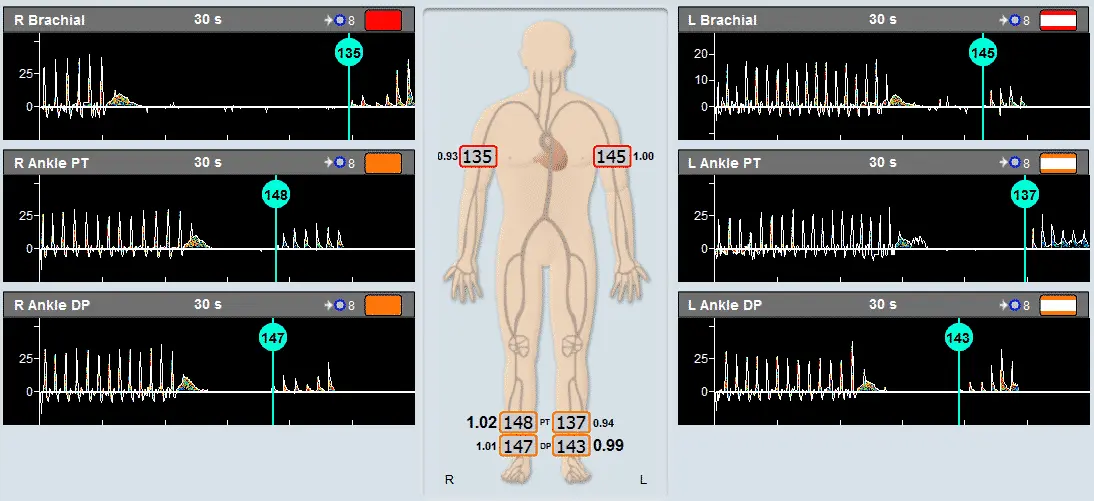
Q: When we perform ABI, our focus is to find the BP based on the waveform and the sound. Should we pay attention to or report the amplitude of the signal?


A: When perform ABI, it is important to focus on obtaining the best Doppler signal in order to accurately measure BPs.


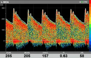
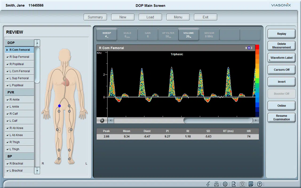
Spectral Doppler waveform assessment is a diagnostic tool used in the diagnosis of peripheral vascular disease. This consensus statement from the Society for Vascular Medicine and Society for Vascular Ultrasound gives useful information on interpretation of peripheral arterial and venous Doppler waveforms (https://doi.org/10.1177/1358863X20937665).


image1 31


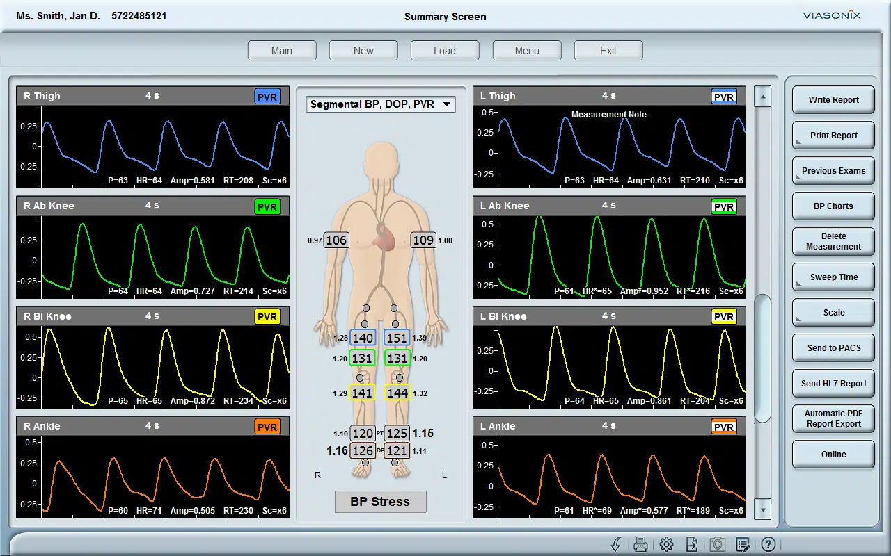
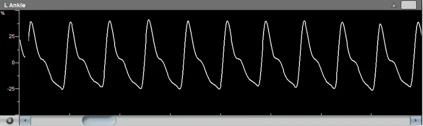
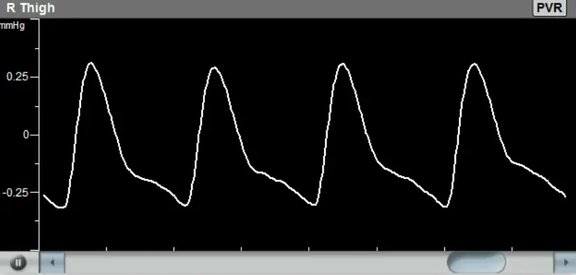
Q: When we perform PVR, should we report the waveform of the PVR or how to describe it in the report? Should we include the amplitude of the signal or the systolic rising time in the report? How reliable of these information?


A: Similar to Doppler waveform, PVR waveform assessment is useful in the diagnosis of peripheral vascular disease. This research report describes how to use PVR waveform analysis to identify inflow disease (https://doi.org/10.1177/8756479314548066).


image2 33


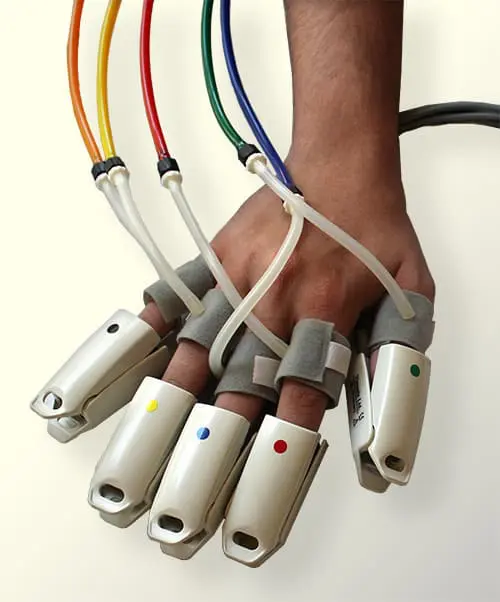
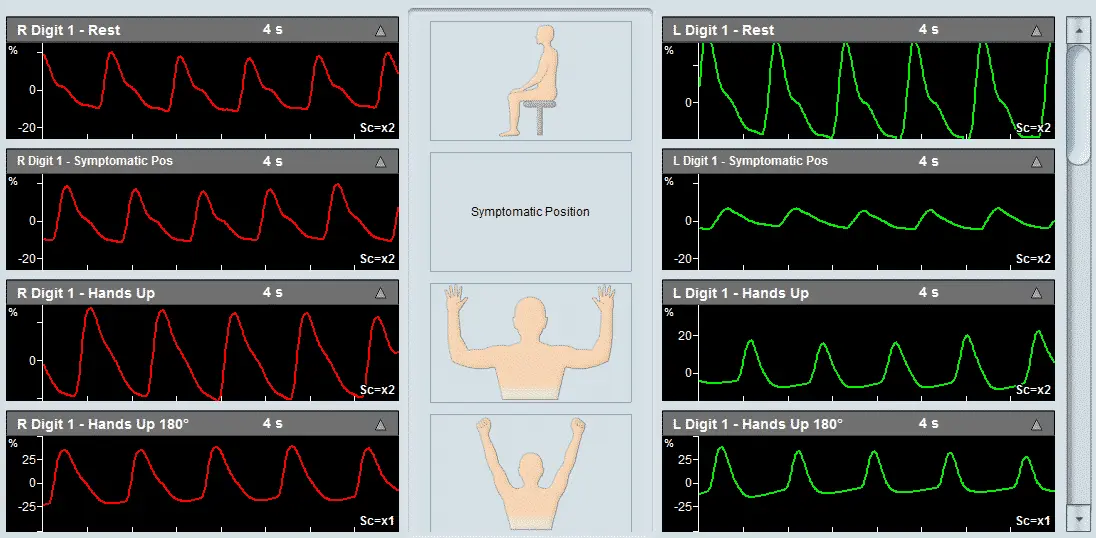
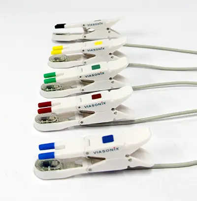
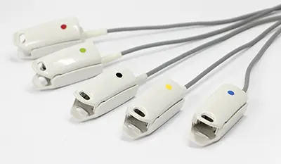
Q: When we perform TBI by using PPG in the dark, we normally focus the BP. Should we report the waveform or how to describe it in the report? Should we include the amplitude of the signal or the systolic rising time in the report? If the patient with 1st and 2nd toes amputated, should we proceed to other toes?


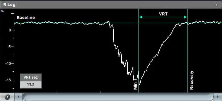
A: TBI is very useful especially when tibial arteries are incompressible. Additional toe waveform analysis may increase the accuracy of assessment for critical limb ischemia (https://doi.org/10.1016/S0741-5214(96)70101-5).


When patients have great toe amputated, you may proceed toe pressure measurement on other toes. However, you should remember results of most published studies on toe pressure are based on great toe. Furthermore, small toes may be too short to apply blood pressure cuff.


image3 35


Q: The reproducibility of the toe waveform’s amplitude is quite poor and very often the amplitude is inconsistent with the ABI. Any suggestions to improve the reliability of the above tests.


A: 1) Every test has its limitations; 2) Practice makes perfect.


image4 37

References:

  1. Interpretation of peripheral arterial and venous Doppler waveforms: A consensus statement from the Society for Vascular Medicine and Society for Vascular Ultrasound (https://doi.org/10.1177/1358863X20937665)
  2. Objective Lower Extremity Arterial Plethysmographic Waveform Characteristics for Differentiating Significant Inflow Disease in Nondiabetic Patients (https://doi.org/10.1177/8756479314548066)
  3. Value of toe pulse waves in addition to systolic pressures in the assessment of the severity of peripheral arterial disease and critical limb ischemia (https://doi.org/10.1016/S0741-5214(96)70101-5)
Read More
As a neurology surgeon at a hospital, I often use Transcranial Doppler (TCD) to track vasospasm, which is a common complication following aneurysmal subarachnoid hemorrhage (SAH). Vasospasm is characterized by a constriction of the blood vessels in the brain, which can lead to decreased blood flow and oxygenation to the brain, and can ultimately result in brain injury or stroke.
Read More

Transcranial Doppler (TCD) is a non-invasive diagnostic tool that measures blood flow velocity in the arteries of the brain. TCD has various applications in neurology and stroke, including:


  1. Detection of intracranial stenosis: TCD can detect intracranial stenosis, which is the narrowing of an artery in the brain. This narrowing can lead to stroke, and early detection with TCD can help prevent stroke.
  2. Diagnosis of vasospasm: TCD can diagnose vasospasm, which is a narrowing of the blood vessels in the brain that can occur after a subarachnoid hemorrhage. Vasospasm can lead to stroke, and early detection with TCD can help prevent stroke.
  3. Monitoring of cerebral autoregulation: Cerebral autoregulation is the ability of the brain to maintain a constant blood flow despite changes in blood pressure. TCD can monitor cerebral autoregulation, which can be useful in patients with traumatic brain injury or after cardiac surgery.
  4. Monitoring of emboli: TCD can monitor emboli, which are small particles that can travel through the bloodstream and cause blockages in the brain. Emboli can be a sign of underlying cardiovascular disease and can increase the risk of stroke.
  5. Detection of right-to-left shunts: TCD can detect right-to-left shunts, which are abnormal connections between the right and left sides of the heart. These shunts can lead to paradoxical embolism and increase the risk of stroke.

In stroke management, TCD has various applications including:


  1. Diagnosis of stroke subtype: TCD can diagnose the subtype of stroke, such as ischemic or hemorrhagic stroke, based on the pattern of blood flow velocity in the brain.
  2. Prediction of stroke risk: TCD can predict the risk of stroke by detecting high-risk features, such as emboli or stenosis, and monitoring changes in blood flow velocity over time.
  3. Monitoring of stroke recovery: TCD can monitor changes in blood flow velocity in the brain as a patient recovers from stroke, which can be useful in assessing the effectiveness of treatment.

In summary, TCD has various applications in neurology and stroke, including the detection of stenosis, vasospasm, and emboli, monitoring of cerebral autoregulation and recovery from stroke, as well as the diagnosis and prediction of stroke subtype and risk. TCD is a non-invasive and convenient diagnostic tool that can be used at the bedside, making it a valuable addition to the clinical management of neurological and cerebrovascular diseases.

Should you require any additional advice, or would like to organise a free demo of our TCD DOLPHIN system, please feel free to reach out to us at MedTech Edge on  info@medtechedge.com  and we assist you with the right TCD product for your practice/department or research project.

image1 1

Read More

Transcranial Doppler (TCD) is a non-invasive diagnostic tool that uses ultrasound waves to measure blood flow velocity in the arteries of the brain. MRI (Magnetic Resonance Imaging) is a non-invasive diagnostic tool that uses a powerful magnetic field and radio waves to produce detailed images of the brain.

Here are five potential reasons why TCD might be used instead of MRI:


  1. TCD is less expensive: TCD is generally less expensive than MRI, which can be an important factor for patients or healthcare systems that are cost-conscious.
  2. TCD is faster: TCD is a relatively quick procedure that can be performed at the bedside, while MRI can be a longer and more involved process that may require scheduling and travel to a separate facility.
  3. TCD is more portable: TCD equipment is often more portable than MRI machines, which can make it easier to use in remote or underserved areas.
  4. TCD is better for monitoring: TCD can be used to monitor changes in blood flow velocity over time, which can be useful for tracking the progression of a disease or the response to treatment.
  5. TCD is safer for certain patients: TCD is a non-invasive procedure that does not expose patients to ionizing radiation, which can be a concern for some patients, such as pregnant women or those with a history of cancer.

Screen Capture Main Screen

Read More

Transcranial Doppler (TCD) technology has been rapidly advancing in recent years, allowing for non-invasive monitoring of cerebral blood flow. Here are some of the latest advances in TCD technology:


  • Automated emboli detection: Modern TCD machines have automated emboli detection features that use signal processing algorithms to detect emboli in real-time. This can provide more efficient and accurate detection of emboli, which can be important in diagnosing and monitoring cerebrovascular disease.
  • Multi-gate technology: Multi-gate TCD technology uses multiple probes to detect blood flow velocity at various depths within the brain, providing more detailed information about blood flow patterns in the brain.
  • TCD robotics: leading edge TCD systems incorporate robotics to assist the user in quickly and automatically insonating and monitoring cerebral blood flow.
  • Wireless connectivity: Some newer TCD machines offer wireless connectivity, allowing for seamless integration with electronic medical records and other hospital systems.
  • Artificial intelligence (AI) and machine learning: Some TCD machines are now incorporating AI and machine learning algorithms to aid in the interpretation of TCD data. This can help to improve accuracy and efficiency in diagnosing and monitoring cerebrovascular disease.
  • Portable devices: Some TCD machines are now more compact and portable, allowing for easier use in clinical and research settings where space is limited or where patients cannot easily be moved to a larger machine.

Overall, these advances in TCD technology are improving the accuracy, efficiency, and convenience of non-invasive monitoring of cerebral blood flow.

Should you require any additional advice, please reach out to us at MedTech Edge on info@medtechedge.com and we will assist you with the right TCD product for your practice/department or research project.

dolphin 4d

Read More