What are Vascular Doppler Measurements?
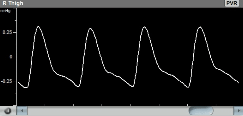
Doppler measurements of the peripheral arteries, particularly those in the legs, are an important qualitative diagnostic method to determine the arterial vascular elasticity, as part of a complete Peripheral Arterial Disease (PAD) diagnosis.
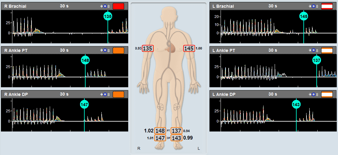
The primary assessment includes the determination of the number of “phases” in a Doppler waveform over one cardiac cycle, typically between one (abnormal) and 3 (good elastic vessel).
In the lower extremities, the arteries that are typically measured with Doppler include the Dorsalis Pedis, Posterior Tibial, Popliteal, and Femoral arteries. In the upper extremities, the most commonly measured arteries include the Radial, Ulnar and Brachial vessels.
Which Doppler Probe should be Used?
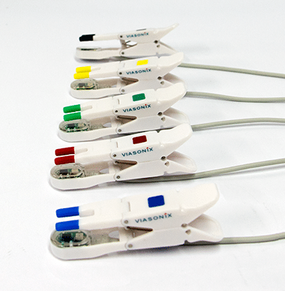
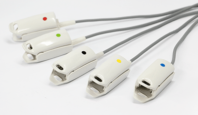
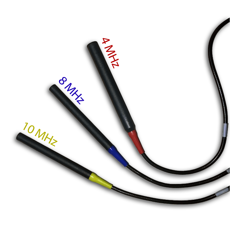
The requirements from the Doppler probe is to have an appropriate frequency that meets the measurement requirements. Thus, for the Doppler measurement of the smaller, more superficial vessels such as the DP or PT, a 10 MHz or 8 MHz probe is used, while for larger or deeper vessels, the 4 MHz probe is preferred.
Also, the requirement from the Doppler is to have Continuous Wave (CW) Doppler, direction sensitivity and bi-directional flow capabilities, audible output, and recording. Above all, a complete color spectral analysis is a preferred option as it allows us to distinguish between forward and reverse blood flow clearly.
Doppler Probes
Gold Standard ABI Measurement Method
Using the Falcon for Doppler Measurements
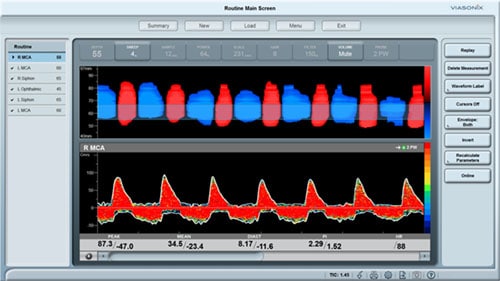
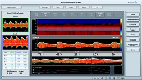
The Falcon provides a wide range of CW Doppler probes that cover the complete range of measurements in peripheral arteries. These probes include the standard 4 MHz and 8 MHz probe frequencies, as well as a 10 MHz probe for very shallow and small vessels. Unlike many other systems in the market, the Falcon provides complete spectral analysis in full color, which allows to clearly distinguish between forward and reverse flows. This helps preventing the inaccuracies of displaying just an envelope which could be mistaken as just noise, or vice versa.
All Doppler settings can be pre-configured in the protocol, so in order to complete the test only a single button can be used. The user can modify the preferred sweep time display, scale, gain, filter or sound volume. Many other Falcon features and options are designed to simplify the use of the Falcon Doppler physiologic measurements followed by a complete clinical diagnosis, in a fast and efficient way.
While the diagnosis of the Doppler measurements is primarily qualitative, the Falcon also provides a range of quantitative parameters, including the Peak, Mean and Diastolic velocities, the PI, RI and S/D pulsatility indices, and the rise time and heart rate. These parameters allow for improved quantification and diagnosis of a primarily qualitative test.
Expected Results
The diagnosis of the peripheral Doppler waveforms is primarily qualitative in nature. The focus is on determining the number of “phases” in the Doppler signal. If the waveform has a dominant forward flow, followed by a smaller reverse flow section and then another small forward flow, this waveform is considered to have 3 phases or a tri-phasic waveform. Likewise, if the final small forward flow is not seen, then the signal is considered bi-phasic, and if only the initial forward flow is seen, then the signal is considered mono-phasic.
Typically, a “normal” Doppler waveform is a tri-phasic waveform, indicative of good and elastic arteries. With the progression of arterial disease, the vessels become stiffer, and the third and then the second flow phases start to disappear. With a monophasic Doppler waveform, the systolic peak is also less sharp and becomes wider and rounded.

