What is Venous Reflux?
The Venous Reflux test is used to determine the competence of the superficial venous valves in the calves of the legs. This test is performed with a DC PPG sensor.
When the venous valves are not functioning properly, they may “leak”, resulting in a reversal of blood flow in the veins. In turn, this problem causes improper venous blood flow and pooling of blood in the veins of the legs, which may result in a variety of venous insufficiency disease. The Venous Reflux test is a common non-invasive diagnostic examination designed to determine the venous valves’ competence in the lower part of the legs.

How to Perform Venous Reflux Test
During a typical Venous Reflux examination, the patient is requested to sit upright. In some protocols, the patient’s feet do not touch the floor, while in other protocols, the feet are flat on the ground. A Photo-plethysmograph (PPG) sensor is attached to each leg above the ankle in the posterior tibial artery region, and the patient is asked to sit still without moving the legs.
Once a steady baseline DC PPG signal is obtained, the patient is asked to perform multiple rapid leg dorsiflexions resulting in “pumping” and emptying all of the venous blood in the upward direction. When the feet are in the air and off the ground, both dorsi- and plantarflexions of the feet are possible for more effective emptying of the venous blood in the calf. The number of dorsiflexions varies between institutes and is frequently 10.

During the dorsiflexions of the feet, the venous blood in the veins is driven in the proximal direction towards the heart, and this causes the PPG signal, which represents the blood in the veins, to drop sharply. Immediately after completing the dorsiflexions of the feet, the patient is requested to remain still without moving the feet to allow to refill the veins in the legs with blood. As a result of the refilling of the veins, the PPG signal returns back towards the initial baseline.
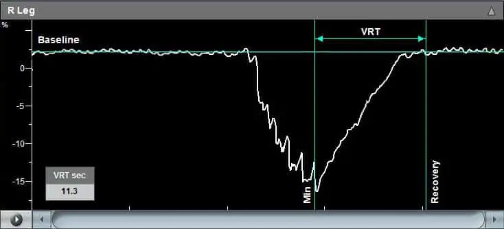
The duration from the end of the dorsiflexions (maximal PPG signal drop) and until the signal return to baseline is called the Venous Recovery Time (VRT), which is a strong indication of the status of the valves. A slow recovery time indicates competent valves, while a fast VRT suggests a suspicion of possible incompetency of the venous valves.
If the venous reflux test indicates suspicion of venous insufficiency, then a venous tourniquet can be applied at various positions above or below the knee to discriminate between deep and superficial valve incompetency.

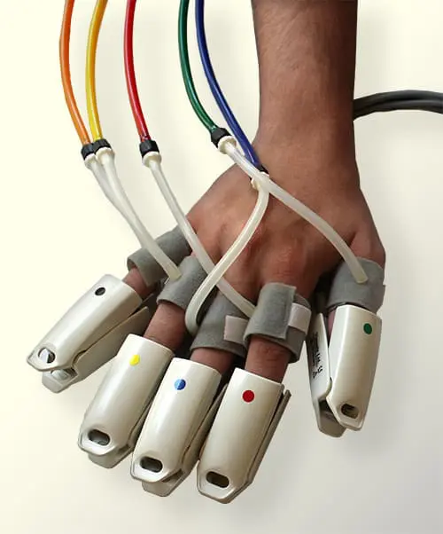
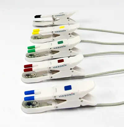
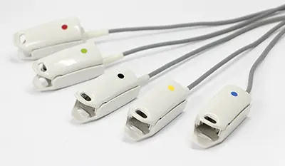
PPG Sensors
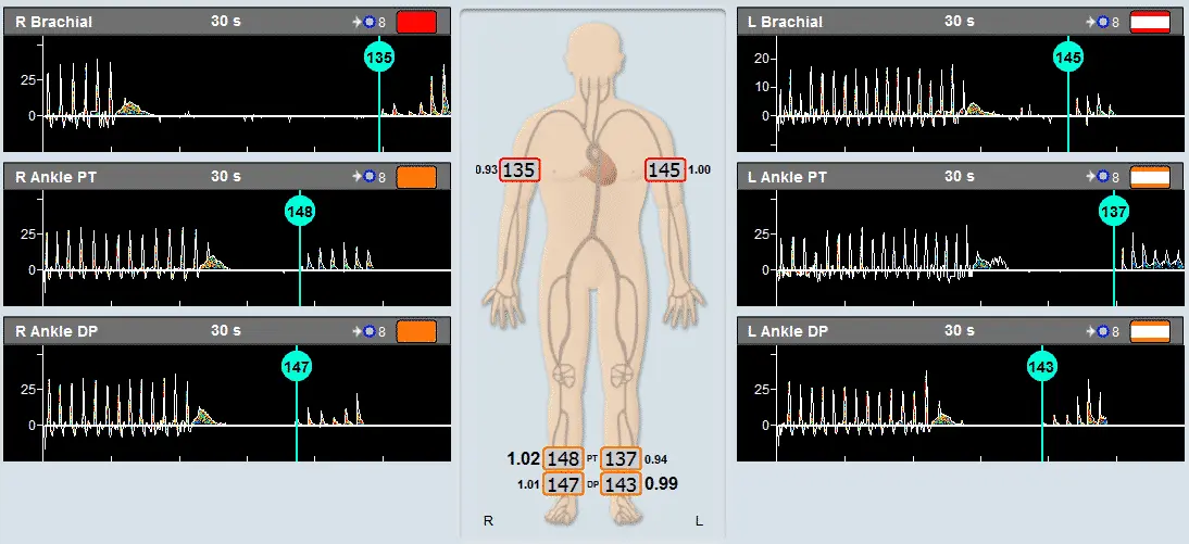
Secondary Method of ABI Assessment
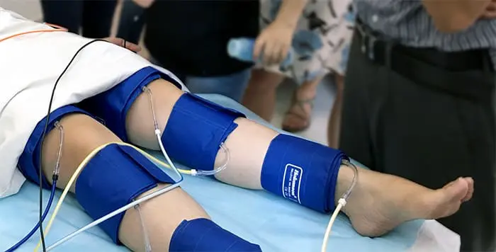
Inflatable Cuffs
High quality available in a variety of sizes
Using the Falcon for Venous Reflux Test
The Falcon is designed with a dedicated Venous Reflux Test protocol to allow a fast, simple, and effective diagnosis. The Falcon disk PPG sensor is attached using a special double-sided adhesive sticker to the proper location on the lower calf. The Falcon Venous Reflux protocol then guides the examiner through the simple steps that need to be taken to complete the test effectively. These steps include:
- Obtaining a steady-state PPG signal,
- Placing an automatic signal “baseline” cursor
- The patient starts the vigorous dorsiflexions and/or plantarflexions.
- Once the vigorous foot movements are stopped, and the patient is still again, the PPG signal starts to rise towards the baseline, and an automatic time-cursor is placed at the point of minimal PPG signal value.
- Finally, once the drifting signal crosses the initial baseline value, the “Recovery” time cursor is placed at that location. The VRT parameter – Venous Refill Time – is automatically displayed as the time difference between the minimum PPG signal and the point of recovery.
The examiner or physician has the option to relocate and move the time cursors to adjust the VRT parameter per their understanding or in the event that the PPG signal did not fully recover back to baseline.
The diagnosis of the Venous Reflux test is quantitative and is based on the value of the VRT parameter.
The Falcon allows to extend the VR test protocol based on the VRT findings and add a tourniquet pressure cuff that is placed on the leg, typically above and below the knee. The examiner can set the tourniquet venous occlusion pressure. The venous reflux test can be repeated with the tourniquet cuff to determine deep or superficial valve incompetency.
Expected Results
The diagnosis of the Venous Reflux test is quantitative and is based on the value of the VRT parameter. Typically, a VRT value greater than 20 seconds is considered normal, while a VRT value lower than 20 seconds indicates venous reflux and possible venous valve insufficiency.
The application of venous tourniquet aid with the discrimination between deep and superficial valve insufficiency. If an abnormal VRT value becomes normal with the application of a superficial tourniquet, this may indicate that isolated superficial reflux is present. However, if the VRT value does not improve, then either deep venous reflux is present, or both superficial and deep venous reflux is present.
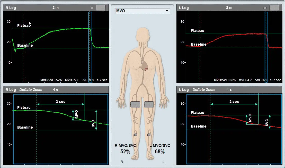
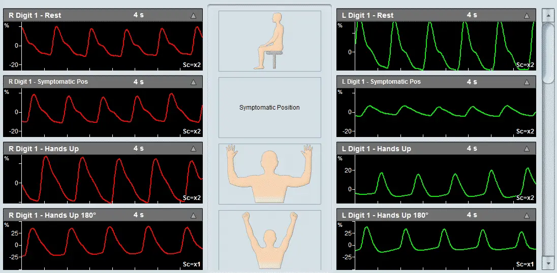
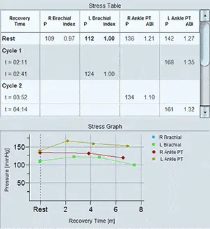
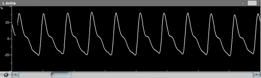
Example of a Venous Reflux specialty test performed on a patient with Viasonix Falcon/PRO. The Venous Recovery Time (VRT) is an index of venous valve patency. A fast recovery time such as in this example, indicates a suspicion of valvular incompetency.