MedTech Edge
MedTech Edge
  • Home
  • About Us
  • Transcranial Doppler
    • Dolphin 4D
    • Dolphin IQ
    • Dolphin Max
    • Dolphin/XF
    • TCD Demo
  • Vascular Diagnostic Systems
    • Falcon Pro
    • Falcon Demo
    • Air Plethysmography
    • Ankle Brachial Index (ABI)
    • Doppler Measurements
    • Exercise Stress Test
    • Extracranial Examination
    • Maximum Venous Outflow / Segmental Venous Capacitance (MVO/SVC)
    • Palmar Arch Test and AV Fistula
    • Penile Function
    • Photo-Plethysmography Readings
    • Pulse Volume Recording
    • Pulse Wave Velocity
    • Raynaud’s Syndrome
    • Reactive Hyperemia
    • Segmental Blood Pressures
    • Thoracic Outlet Syndrome
    • Toe Brachial Index (TBI)
    • Venous Reflux
  • Vascular Doppler Ultrasound
  • ABI Machines
  • PAD Testing
  • Contact
  • Blog

404

  1. Home
  2. 404

ERROR 404
–
NOT FOUND

You may have mistyped the URL,
Or the page has been removed

<< Go back to home page >>

LOOKING FOR A LEADING EDGE SOLUTION FOR YOUR NEUROLOGY / TRANSCRANIAL DOPPLER LABORATORY?

ASK OUR EXPERTS

Contact Us
  • Home
  • About
  • Blog
  • Contact
  • Sitemap

Contact Us

mob: +61 466 060 220
info@medtechedge.com

Level 1, 54 Davis Avenue
South Yarra 3142
VICTORIA AUSTRALIA

Partners

MedTech Edge – all rights reserved 2013-2023

Sickle Cell Disease

Sickle cell disease is a disorder of the blood caused by abnormal hemoglobin which causes distorted (sickled) red blood cells. It is associated with a high risk of stroke, particularly in the early years of childhood.


Transcranial Doppler (TCD) can be significant in the prevention of stroke under this condition. Mean of maximum cerebral velocity readings are obtained, and results are classified as normal, conditional, abnormal or inadequate based on the measurements. Clinical treatment based on the findings can be significant in stroke prevention. 


The Viasonix Dolphin is ideal for sickle cell disease diagnosis and monitoring, with a dedicated sickle cell protocol. Special markers, which can also be configured by the examiner, clearly mark the velocity thresholds according to international guidelines, help in the identification and documentation of the pathology.

Brain Death

Brain Death condition reflects an extensive and irreversible brain damage, characterized by almost no volume blood flow to the brain. Timely identification of brain death is important in cases of organ donations.
TCD can detect the different stages of brain death through analysis of the particular waveforms: a sharp systolic peak followed by near zero diastolic velocities; systolic spikes followed by retrograde diastolic velocities; short spikes; and ultimately, no signals at all. Note that there are countries in which the use of TCD for diagnosis of brain death is not accepted.


For determination of brain death, the characteristic waveforms should be displayed in all intracranial vessels. In order to accept the absence of a Doppler signal in any vessel, a prior valid measurement must be identified in the same vessel to ensure that the access temporal window is fine.


The Viasonix Dolphin special m-mode option allows the examiner to view flows at all depths along the ultrasound beam. Thus, even after the measurement is freezed the examiner can scroll back and view signals from any depth and replay the spectrum from any depth, even if it is different than the original set depth. This significantly reduces the risk of missing a valid blood flow signal and reaching an erroneous clinical diagnosis.

Collateral Capacity

Similar to cerebral autoregulation and vasomotor reactivity, cerebral collateral flow reflects an alternative compensatory mechanism to pathological flow conditions. The cerebral collateral pathways include side-to-side via the anterior communicating artery, posterior-to anterior via the posterior communicating artery, and external-to-internal via the ophthalmic artery. TCD can detect reverse velocities in the collaterally perfused arteries, indicating the viability or absence of this compensatory mechanism.


The Viasonix Dolphin’s highly sensitive M-mode display identifies forward or reversed blood flow, throughout the depth range, for analysis of this brain protection mechanism.

Patient Foramen Ovale (PFO)

Transcranial Doppler is very useful in the identification of patent foramen ovale (PFO). PFO is a hole between the left and right atriums of the heart. While this hole exists in everyone before birth, it most often closes shortly after birth. If the hole fails to close naturally after birth it is called PFO.


TCD can easily identify the existence of PFO by injecting micro-bubbles (air mixed saline) to the patient vein (typically the antecubital vein), while the Doppler signal is recorded during the Valsalva maneuver. If embolic events are found in the recorded cerebral blood flow, typically the middle cerebral artery (MCA), then this is an indication of the existence of PFO in the patient.


The Viasonix Dolphin is ideal for PFO examinations, with a dedicated PFO protocol. The test can be performed unilaterally or bilaterally, with automatic count of high intensity transient signals (HITS) which are considered as embolic events. Each embolic event can be further analyzed with extremely high temporal and depth resolutions, and the specifications of each event displayed in a special screen. Furthermore, the embolic travel in the depth and time domains can be viewed for additional discrimination from artifact events.

Emboli Detection

Cerebral emboli can take the form of atherosclerotic plaques, air bubbles or blood clots. The immediate risk of cerebral embolization is in impaired perfusion which may lead to stroke and death.


TCD can detect the embolic signals in the blood flow velocity waveforms, which are characterized by high intensity, short duration and unidirectional signals. High intensity transient signals (HITS) are often considered as embolic events.


The Viasonix Dolphin has the most advanced HITS detection and analysis platform. Dedicated screens show the specific embolic characteristics, such as its’ energy, velocity and duration patterns, as well as display its’ travel route in the time and depth domains.


Furthermore, the Dolphin can detect embolic events that are even not within the measured spectrum of the central depth, due to its’ powerful multi-range and multi-depth analysis capabilities.

Monitoring

Cerebral monitoring can be critical for identification of neurological complications. Relative and/or sudden changes in cerebral blood flow velocities can become a powerful diagnostic tool and effect the immediate clinical intervention.


Bilateral cerebral monitoring is particularly useful during cardiac surgery for identification of embolic events, which may be the source of post-surgery neurological deficiencies.


The Viasonix Dolphin supports both unilateral and bilateral cerebral blood flow monitoring with a dedicated and easy to use monitoring headset. A variety of monitoring tools allow to view the complete duration of the monitored procedure, as well as zoom in or out on specific regions of interest. A variety of automatically inserted event markers, as well as user defined markers, help in post-monitoring analysis.


In addition, the Dolphin supports the display of data obtained from external channels for close correlation with the cerebral blood flow information.

Breath Holding

The Breath Holding (BH) test is a specific test for evaluation of Vasomotor Reactivity (VMR) and cerebral autoregulatory capacity.


Identification of exhausted VMR can be a good indication for high risk for stroke. Breath holding induces and increase in CO2, which in turn results in cerebral vasodilation. An increase in peak velocities during breath holding indicates different levels of patent autoregulatory capacity, While the absence of flow increase during the BH test indicates diminished physiological vasodilatory capacity and potentially high risk for stroke.
The Viasonix Dolphin is ideal for BH test with a dedicated breath holding protocol. This protocol leads the examiner with the different stages of the test, and provides automatically the breath holding index (BHI) for clinical determination.


A large timer displayed during the test helps both the patient and the examiner in keeping track of the duration of the procedure. Automatically placed markers identify the peak and minimal velocities measured during the procedure.

VMR and Autoregulation

Vasomotor reactivity (VMR) and autoregulation define the physiological compensatory mechanisms when flow is impaired. Identification of exhausted VMR can be a good indication for high risk for stroke. VMR can be assessed with TCD under two specific conditions: hypercapnia or CO2 reactivity, and with intravenous injections of Acetazolamide or the Diamox test. Note that the Diamox test is not accepted in some countries.


Typically, an increase in peak velocities above 40% during induced increase in CO2 reflects intact VMR, whereas a smaller increase reflects different degrees of impaired VMR. Absence of flow increase reflects a complete exhaustion of the autoregulatory capacity.


The Viasonix Dolphin is ideally set with dedicated Unilateral and Bilateral protocols to determine the autoregulatory capacity of the cerebral circulation of the patient. Doppler waveforms are captured automatically during baseline, and peak (hypercapnia) and minimal (hypocapnia) flows for immediate evaluation, as well as a display of the blood flow fluctuations during entire test process. The respective parameters are automatically shown in a dedicated table with the VMR index readily displayed.

Intracranial Stenosis

Intracranial stenosis relates to a focal narrowing of the artery, either as a result of an atherosclerotic lesion or as a result of external compression due to a tumor.


TCD will easily identify a local increase in blood flow velocities, characterized by the fact that proximal or distal to the stenosis the velocities will be much lower that at the site of lesion itself. Peak focal velocities above 150 cm/sec can indicate an arterial stenosis. However, if the severity of the stenosis is beyond its’ critical level, velocity signals may be diminished. 


The Viasonix Dolphin ideally allows multi-depth scanning along the ultrasound beam, and display in both the m-mode and spectral domains. This allows for simultaneous analysis and evaluation of pre- and post-stenotic flows as well as in the focal region of the stenosis itself.

Evaluation of ICP Changes

Intracranial pressure (ICP) is the pressure inside the skull and thus in the brain tissue and cerebrospinal fluid. Elevated ICP has a direct effect on the cerebral blood flow velocity and its’ pulsatility, which can be detected with simple transcranial Doppler (TCD) measurements.


The Viasonix Dolphin has the ability for multi-parameter analysis which allows close evaluation of cerebral condition. In addition, dedicated trend analysis displays help to closely monitor changes in cerebral condition for timely identification of deterioration of cerebral perfusion.

Alteriovenous Malformation

Arteriovenous Malformation (AVM) relates to a condition whereby the arterial flow bypasses the capillary circulation and connects directly to the venous drainage, thus not allowing for appropriate tissue perfusion.


This condition is characterized by low resistance to blood flow, resulting in high volume flow rates. AVM is therefore characterized by high mean velocities – although lower then with severe vasospasm – which are coupled with a low pulsatility index (PI).


The Viasonix Dolphin multi-depth options allows to simultaneously view and measure the blood flow spectrum at different depths along the beam and quickly identify deviations from expected waveform patterns.

Vasospasm and SAH

Cerebral Vasospasm is typically the outcome of Subarachnoid Hemorrhage (SAH) as a result of an aneurysm rupture. Early detection of vasospasm is critical since it allows timely intervention.


TCD is rather specific for this condition, and allows non-invasive, continuous bed-side monitoring.  Vasospasm is characterized by increased velocities due to arterial narrowing. Severe vasospasm is defined in various international guidelines, and typically when mean velocities exceed 200 cm/sec, moderate conditions are with mean velocities between 120-200 cm/sec, and mild vasospasm is for mean velocities near 120 cm/sec. Additionally, velocities in the middle cerebral artery can be compared to internal carotid artery velocities, and the resulting ratio (Lindergaard index) can indicate cerebral vasospasm when it is greater than 3.


Typically, vasospasm is a developing pathology, and requires day by day monitoring of blood flow velocity values.  The Viasonix Dolphin system provides a dedicated vasospasm protocol that allows setting velocity thresholds for immediate identification of the severity of the vasospasm condition.  In addition, an immediate velocity trend analysis is provided in order to help identify the critical time whereby intracranial velocities are dramatically increased, and immediate clinical intervention is required.


The Dolphin also provides an ideal platform for the identification of the critical highest intracranial velocities.  All data is saved both in the time domain and at all depths along the ultrasound beam, which allows the examiner to review the data after freeze to explore the measured blood flow velocities.

Air Plethysmography

Air Plethysmography (APG) test relates to the use of a pressure cuff as a sensor. The pressure cuff is inflated to a low pressure, such as around 20 mmHg for lymphatic occlusion or around 60 mmHg for venous occlusion, and then the resulting average cuff pressure serves as the sensing element and identifies limb changes.
APG is not a very popular test, because the use of the popular PPG sensors is very simple and easy to use, and is considered in many places the gold standard. Yet, the use of APG is found in places where PPG is not considered reliable enough.
The most popular application for APG is for the venous reflux test.

Extracranial Examination

Extracranial examination is performed with Doppler measurements in the carotid and other extracranial vessels. These measurements focus on the measurement of peak and mean blood flow velocities in order to identify a vascular obstruction to flow, such as an arterial stenosis.
The common blood vessels during extracranial evaluation are the common, internal and external carotid arteries, as well as the subclavian artery. A measurement at the location of a stenosis will result in increased blood flow velocities. 

Palmar Arch Test

The Palmar Arch Test (PAT) is a specialty test performed to evaluate the patency of the Palmar arch in the hand prior to radial or ulnar artery harvesting in artery bypass procedures or before the surgical creation of an upper extremity hemodialysis fistula or graft.


Typically, 2 PPG sensors (photo-plethysmograph) are placed on the two extreme digits (digit 1 and digit 5). The FALCON/Pro allows placing even 5 PPG sensors on all five digits. The Palmar Arch Test protocol begins by measuring the resting PPG waveforms, then measuring the waveforms after selected compressions (compression of the radial artery, ulnar artery, or both) and finally measuring the waveforms after the release of the selected compression.


Compression is typically performed by manually compressing the selected artery or arteries. This procedure allows the examiner to evaluate whether the compression results in impeded or loss of blood flow to parts of the hand. Thus, significant reduction or loss of blood flow to areas of the hand suggests that removing that particular artery may jeopardize hand performance if the artery is removed.


Clear markers in the waveforms indicate when artery compression begins, and when the compression is released.

Penile Function

Penile function or impotence test is used for determination of whether penile function disorders are of vascular nature.


The Viasonix FALCON/Pro allows to use a variety of tests, including the simultaneous combination of different tests. Such tests include Doppler arterial measurements in the penile vessels, blood pressure measurements with a penile pressure cuff, and PVR or PPG waveform measurements.


The results of such tests can either identify a penile vascular problem, or eliminate vascular issues as the cause of impotence.

Reactive Hyperemia

The Reactive Hyperemia test is allowed only in certain countries.
Stress testing is frequently used in peripheral vascular diagnosis to differentiate between different vascular disorders, or to determine the functional severity of an arterial stenosis. Systolic pressures are measured before and after inducing vascular stress. While typically the patient is asked to perform a physical exercise such as exercise on a treadmill, sometimes the patients have difficulties in performing exercise.
In special cases, the reactive hyperemia test (RH) can replace the standard exercise tests and induce vascular stress. RH requires to inflate a pressure cuff in order to occlude the blood flow to the limb or segment of interest for up to several minutes, and then rapidly releasing the cuff pressure in order to generate the hyperemic effect.
It is crucial that a professional examiner will be present near the patient at all time, and immediately deflate the pressure cuffs when necessary.

Maximum Venous Outflow/Segmental Venous Capacitance

The MVO/SVC test determines the Maximum Venous Outflow (MVO) and the Segmental Venous Capacitance (SVC).
The patient lies in a supine position with the legs slightly elevated. A venous occluding cuff is placed on the thigh and a Pulse Volume Recording (PVR) pressure cuff is placed on the calve.
Initially, the sensor (PVR or PPG) is measured and a baseline is determined. Then the thigh cuff is inflated to occlude the venous return (around 60 mmHg) and the changes in the sensor signal are recorded. Once a plateau is reached, the occluding cuff is rapidly deflated. The sensor signal also quickly returns back to the initial baseline. The clinician then determines MVO and SVC based on the sensor signal to determine venous functionality.

Raynaud’s Syndrome

Raynaud’s disease is a rare disorder of the blood vessels, usually in the fingers and toes. It causes the blood vessels to narrow when you are cold or feeling stressed.
When this happens, blood can’t get to the surface of the skin and the affected areas turn white and blue. When the blood flow returns, the skin turns red and throbs or tingles. In severe cases, loss of blood flow can cause sores or tissue death.
Primary Raynaud’s happens on its own. The cause is not known. There is also secondary Raynaud’s, which is caused by injuries, other diseases, or certain medicines.

Thoracic Outlet Syndrome

The Thoracic Outlet Syndrome test is conducted in order to determine the vascular role when the patient’s symptoms are indicative of perfusion loss or neurogenic causes.


This procedure tests for intermittent perfusion loss, particularly in the arms and hands.


Either Doppler, PVR or PPG sensors detect normal resting waveforms in the digits or hands and then the patient is instructed through a sequence of positions such as: Hands-up Test, Adson or Scalene Maneuver, Costoclavicular Maneuver, Allen Test, and Provocative Elevation Test.
The clinician tries to identify a position which significantly reduces perfusion to determine whether the symptoms originate from vascular causes.

Exercise Stress Test

In order to differentiate different vascular disorders, or to determine the functional severity of a stenosis, some of the tests described above are repeated after induced stress.
Typically, the patient will be requested to perform treadmill or similar activity, under the supervision of the examiner.
Most of the time the stress testing will include Segmental Blood Pressures, particularly at the ankles and brachials, and sometimes also Pulse Volume Recording (PVR) and CW Doppler measurements.
Segmental pressures are often measured at different post-stress times to determine also the recovery time from the stress effects. The clinician is particularly interested in identifying significant variants from the resting pressure indices and specifically changes in the Ankle-to-Brachial Index (ABI).
On the left side is an example of a Segmental Blood Pressure measurement with induced stress (exercise in this case) performed using Viasonix FALCON/Pro. The table shows blood pressure measurements in pre-exercise and post-exercise conditions. It is common to take multiple measurements (cycles) in timed intervals after performing the induced stress. The stress graph on the left side shows the recovery of each measured segment starting with pre-stress conditions until the last post-stress cycle.

Photo-Plethysmography Readings

The Photo-plethysmograph (PPG) test detects changes in segmental blood volume, in a similar manner to the Pulse Volume Recording (PVR) test. The PPG technology, however, is different than the pressure-sensor based PVR technology.
With PPG, infra-red signals are transmitted to the skin and reflected back to the sensor, therefore PPG waveforms reflect local and rather shallow skin variations in blood flow.
Typical PPG measurement sites are the toes and digits, and interpretation of the signals is in a similar manner to the interpretation of the PVR waveforms.

Venous Reflux

The Venous Reflux test is used to determine the competence of the superficial venous valves in the calves of the legs. This test is performed with a DC PPG sensor.
When performing Venous Reflux test, the patient is requested to sit upright with the legs not touching the ground. A Photo-plethysmograph (PPG) sensor is attached to the leg above the ankle in the region of the posterior tibial artery. When the DC signal reaches a steady state baseline, the patient is requested to perform multiple leg dorsiflexions which result in “pumping” of all of the venous blood. After the dorsiflexions the patient is requested to remain still, while the PPG DC signal returns back to the initial baseline.
Optionally, the test can be repeated with a cuff wrapped around a segment on the leg (thigh, above/below knee) acting as a tourniquet.
The duration from the end of the dorsiflexions (maximal signal change) until the signal return to baseline (VRT, Venous Recovery Time) indicates the status of the valves. A slow recovery time indicates competent valves, while a fast VRT would indicate a suspicion of valvular incompetency.

Pulse Volume Recording

The Pulse Volume Recording (PVR) test is a pneumo-plethysmographic test used for detection of the segmental volume changes in the limb which result from the flowing blood, as a function of the cardiac cycle.
Similar to the Segmental Blood Pressure (SBP) measurements, dedicated pressure cuffs are placed around all limb segments prior to initiating the test.
The cuffs are then inflated to a pressure that would occlude the venous return, yet will maintain the arterial flow un-obstructed. This pressure is typically 65 mmHg.


Once the cuff pressure is stabilized, the waveform signals which reflect the segmental volume changes can be recorded. The clinicians are normally interested in the qualitative shape of the waveforms: a “normal” PVR consists of a rapid systolic upstroke and a rapid downstroke with a prominent dicrotic notch.


With increasing arterial disease the PVR waveform becomes attenuated, the upstrokes and downstrokes are less prominent and the PVR amplitude decreases and ultimately becomes flat.

Ankle Brachial Index (ABI)

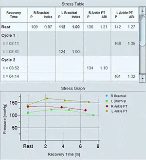
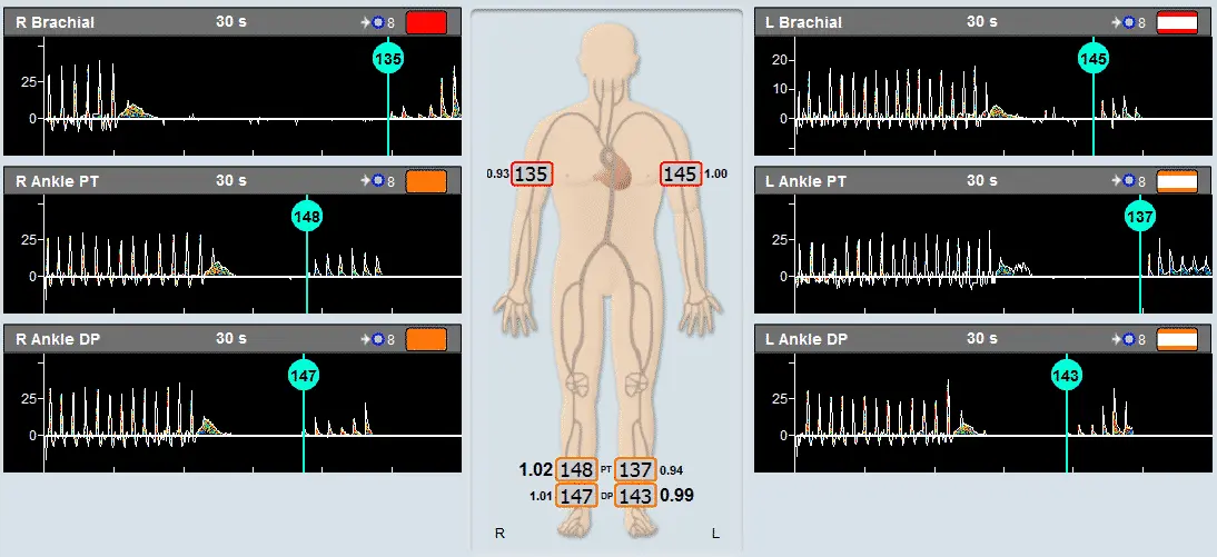
ABI, the Ankle Brachial Index, is the most popular application for fast and simple screening of a physiological vascular pathology. With this test pressure cuffs are placed on the 2 ankles and on 1 or 2 brachial segments.
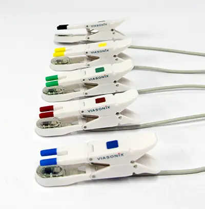
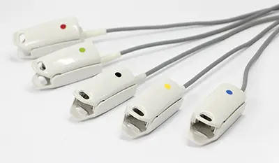
The Viasonix FALCON Pro allows to perform the ABI test with either Doppler or PPG sensors. In addition, the Falcon allows to perform simultaneous measurements in all sites under certain conditions for rapid measurements.
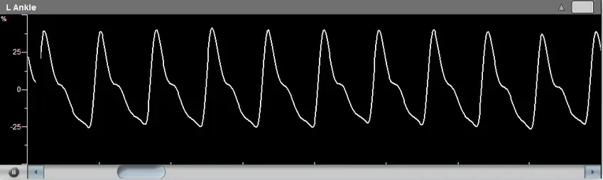
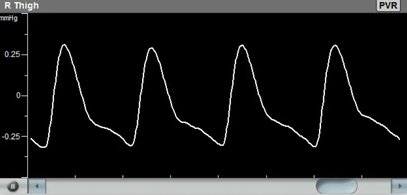
Prior to segmental cuff inflation, a reference signal is identified distal to the cuff location. When the cuff pressure exceeds the systolic pressure, the reference signal waveforms should disappear. Then, the cuff pressure is slowly deflated at constant bleeding rate, and the first occurrence, of the reference signal waveform return, marks the systolic pressure.
The ABI is defined as the ratio of the higher brachial pressure to each of the ankle pressures. A ratio of around 1 is considered normal, while lower values indicate various levels of significant peripheral arterial disease (PAD) according to international guidelines.

Doppler Measurements

Peripheral vascular Doppler measurements are normally performed using CW (Continuous Wave) Doppler probes.
Different Probe frequencies are used depending on the target vessels: 4 or 8 MHz are used for larger and deeper vessels such as the Carotid, Femoral or Popliteal arteries, and higher 8 or 10 MHz are used for the smaller and shallower vessels such as the Dorsalis Pedis or Tibial arteries.
The main objective is to qualitatively examine the waveform shapes: a normal peripheral artery (not in the carotids) will show 3 phases during a cardiac cycle, a prominent early systolic forward flow, a late systolic reverse flow, followed by a small component of forward flow again. This signifies a healthy elastic artery. As the vessel becomes less elastic (for example due to diabetes), the third and/or second phase will disappear.
In addition, significantly high velocities may indicate the presence of a stenosis or some other obstruction to the blood flow.

Segmental Blood Pressure

The Segmental Blood Pressure (SBP) tests require the placement of dedicated pressure cuffs on the various limb segments, which typically include the 2 brachial segments, the thighs (some protocols divide the thigh into 2 separate segments), the segment below the knees, the ankles and the toes.
Additional segments can be measured, such as the digits for diagnosis of the Raynaud’s Syndrome.
Prior to segmental cuff inflation, a reference signal is identified distal to the cuff location. This reference signal is typically a CW Doppler measurement or a PPG (Photo-Plethysmograph, used for the digits and toes).


When the cuff pressure exceeds the systolic pressure, the reference signal waveforms should disappear. Then, the cuff pressure is slowly deflated at constant bleeding rate, and the first occurrence, of the reference signal waveform return, marks the systolic pressure. The clinician is normally interested in the ratio of the ankle systolic pressure to the brachial pressure (ABI – Ankle Brachial Index), or in significant pressure differences either in sequential segments or side-to-side differences for the same segment.