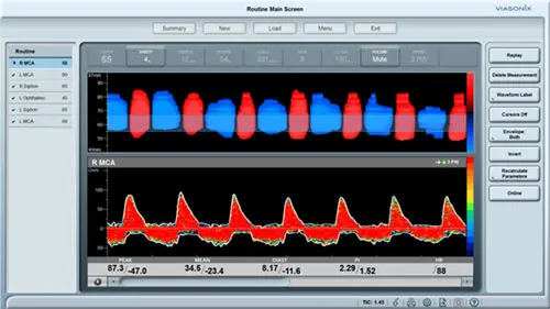
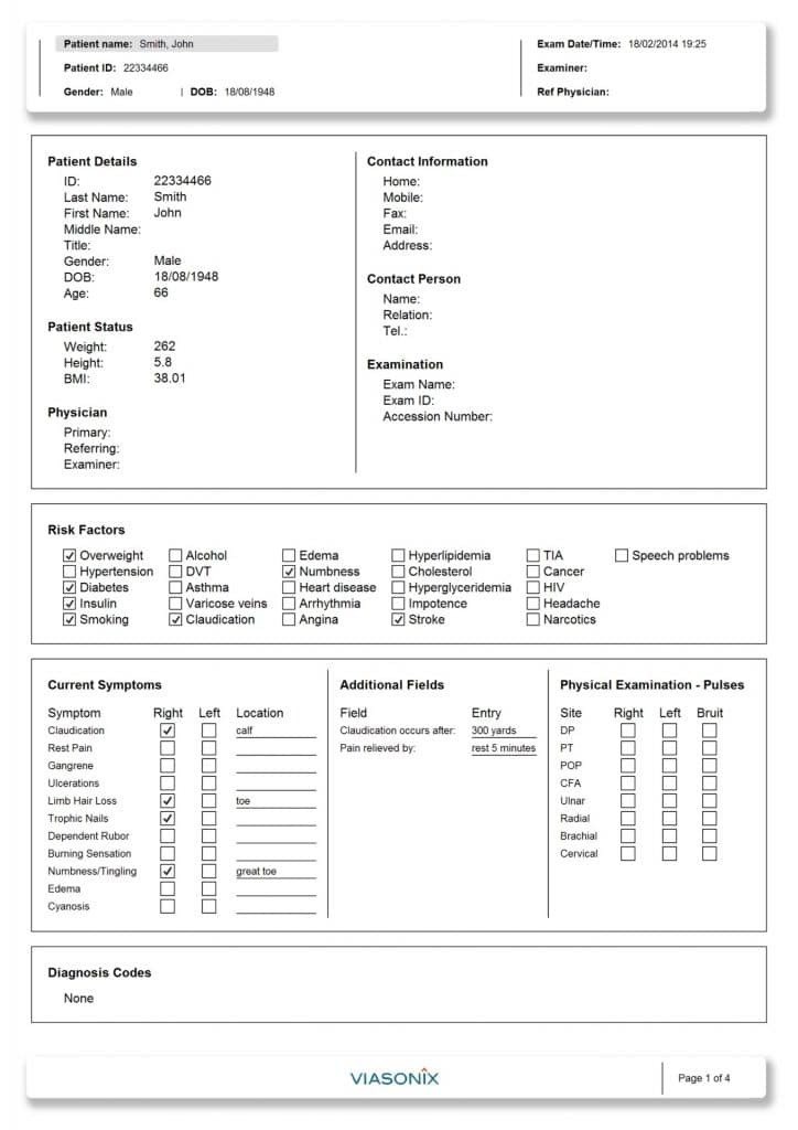
The term peripheral vascular disease (PVD) is commonly used to refer to peripheral artery disease (PAD), meaning narrowing or occlusion by atherosclerotic plaques of arteries outside of the heart and brain. Risk factors for peripheral artery disease include elevated blood cholesterol, diabetes, smoking, hypertension, inactivity, and overweight/obesity
Approximately half of people with peripheral artery disease do not experience any symptoms. For patients with symptoms. Treatments include lifestyle changes, supervised exercise, medications, angioplasty, and surgery.
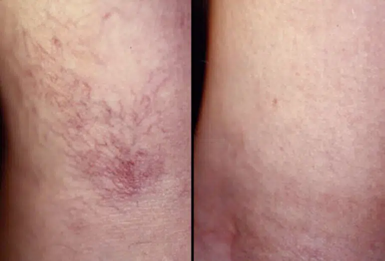
Spider veins and varicose veins are common conditions that affect many adults. These abnormally enlarged vessels, which affect women more often than men, appear most often on the legs and become more prevalent with age.

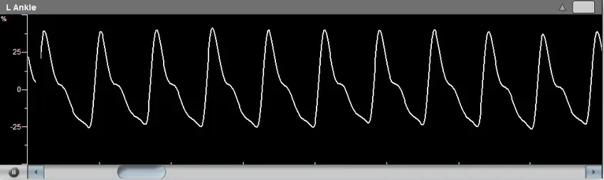
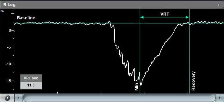
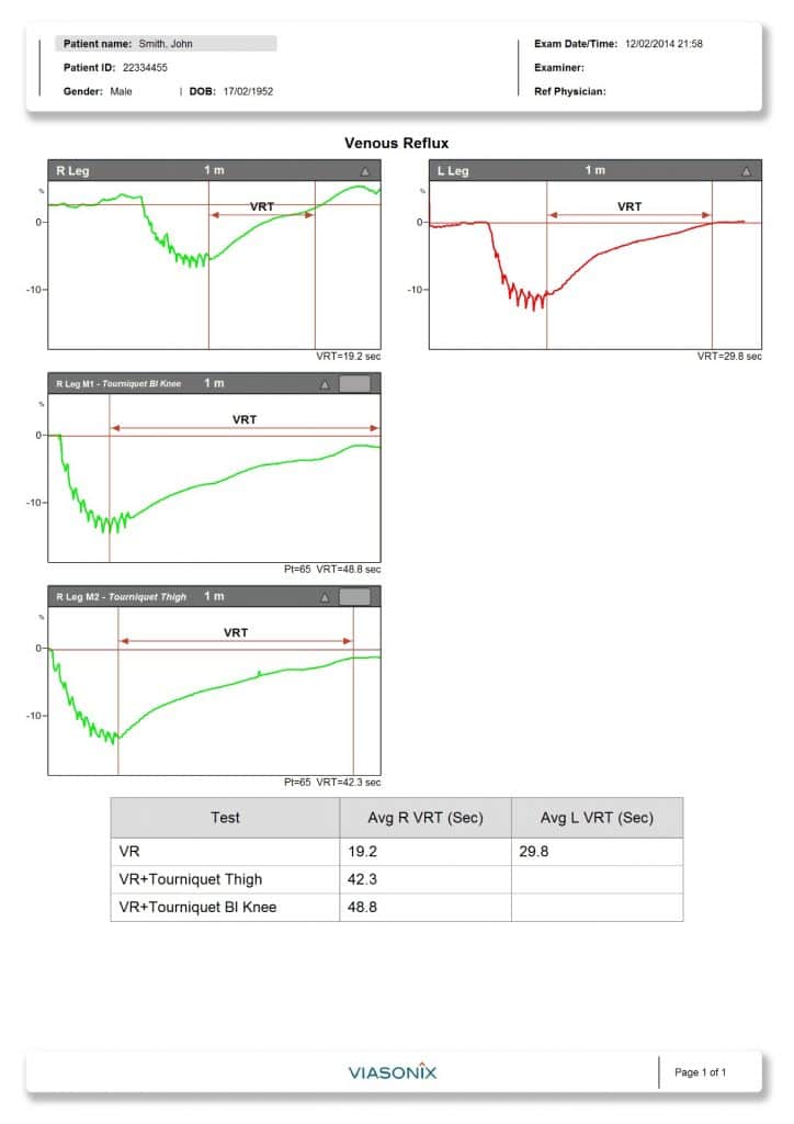
The venous reflux test is designed to assess lower limb venous competence. The test is performed with the patient in a sitting position with the legs in the air (not touching the ground).
To perform the test, place PPG sensor on the patient calf with the aid of the Viasonix adhesive sticker, record PPG signal until the PPG signal level reaches a plateau, ask the patient to perform foot dorsi-flexions on the tested side for several times and request the patient to continue sitting without movement until the PPG signal returns to the baseline level. Falcon Pro will automatically calculate and display Venous Refill Time (VRT) which is recovery time from the lowest point of the PPG signal (after dorsiflexion) to the signal returns to the baseline.
Figure shows right leg has a fast recovery time (short VRT), indicating venous incompetence. A longer VRT at repeated tests with tourniquet suggests venous reflux affects superficial venous system only.

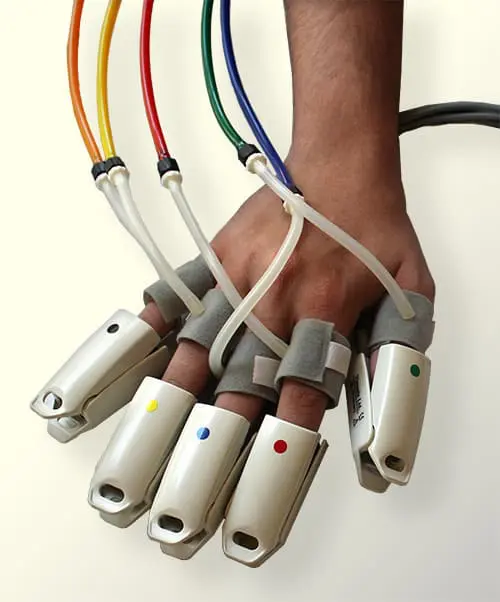
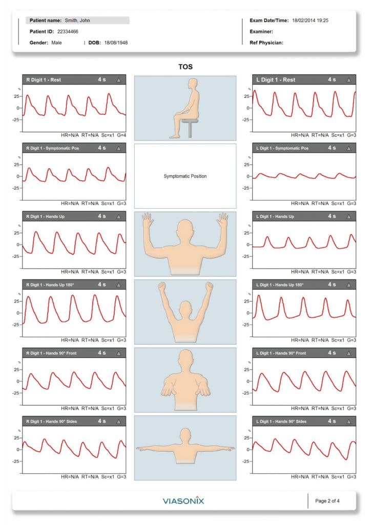
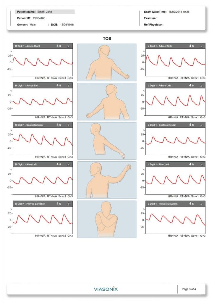
The Thoracic Outlet Syndrome (TOS) test is designed to test an intermittent loss of perfusion to the patient finger or arm during rest and provocative maneuvers. The test may be performed with a photo-plethysmography (PPG) sensor, PVR cuff sensor, or a Doppler measurement. The test is performed with the patient in a sitting position.
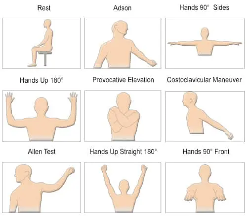
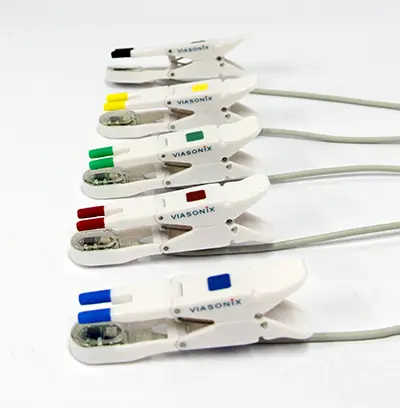
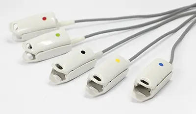
The specialty tests are supported with dedicated pictures that guide the examiner through the examination protocol, and the corresponding reports clearly correspond the measured waveforms to the different protocol steps.
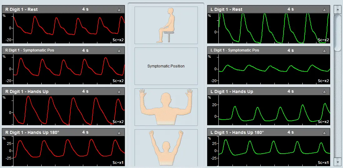
Figure 1 (Page 2) shows bilateral thumb PPG waveforms with arm in positions of rest, symptomatic, hand up, hands up 180°, hands 90° front, hands 90° sides.
Figure 2 (Page 3) shows bilateral thumb PPG waveforms with arm in positions of Adson right, Adson left, costoclavicular, Allen left, and provocative elevation.
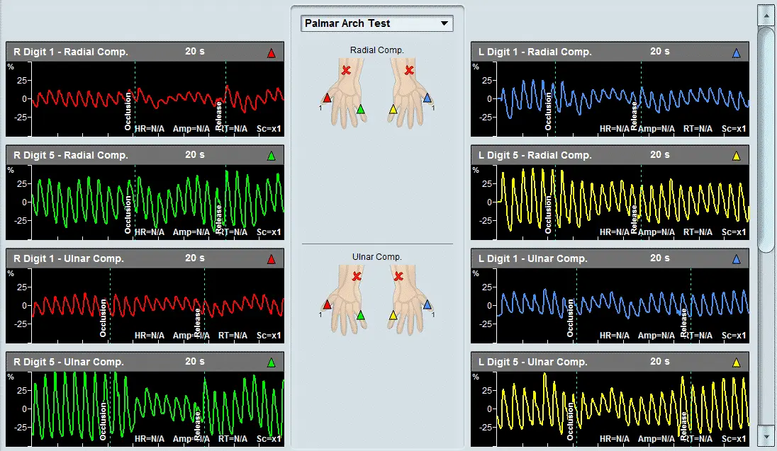
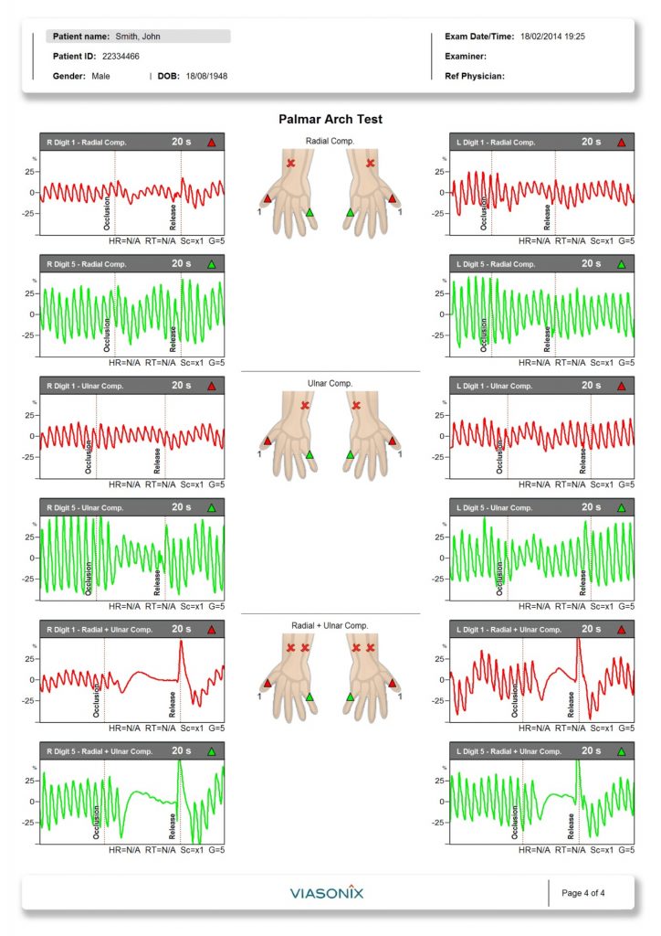
The Palmar Arch test is designed to test the patency of palmar arch prior to harvesting radial artery for coronary bypass or creation of arteriovenous fistula for haemodialysis. The test compares finger PPG waveforms pre and post occlusion of radial artery, occlusion of ulnar artery, and occlusion of both radial and ulnar arteries. The test is performed with the patient in a sitting or supine position.
Figure 3 (Page 4) shows bilateral thumb PPG waveforms pre and post radial artery occlusion (top), bilateral 5th finger PPG waveforms pre and post radial artery occlusion (middle), and bilateral thumb PPG waveforms pre and post ulnar artery occlusion (bottom).
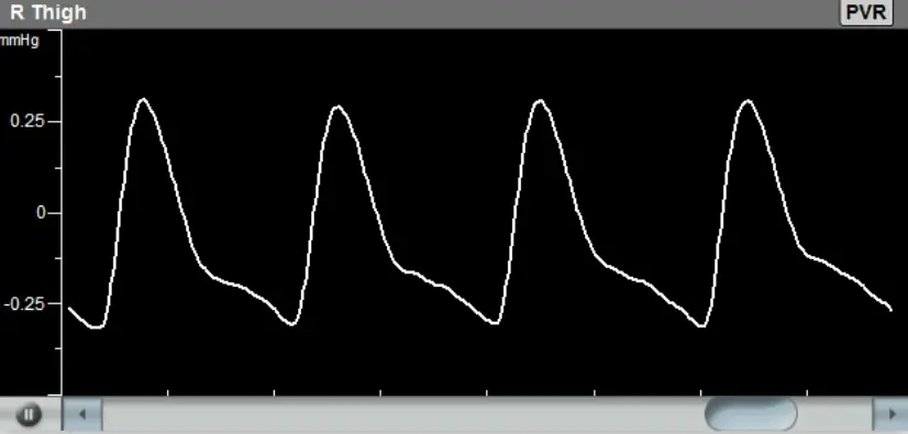
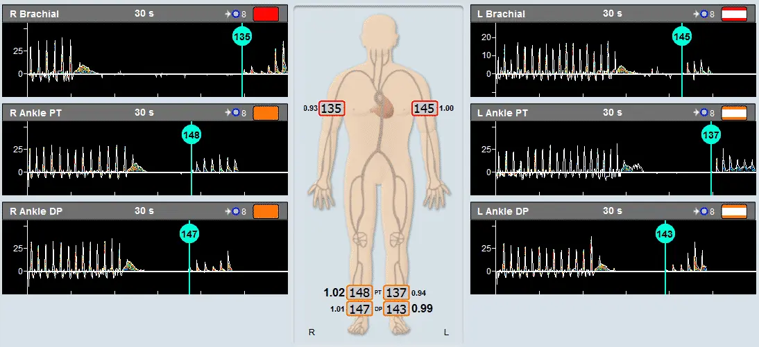
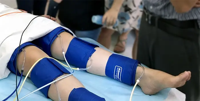
Segmental Blood Pressure Measurement, Doppler Waveform Record, Pulse Volume Recording (PVR), Exercise PVR and Exercise Ankle Brachial Index (ABI) are designed to assess location and severity of lower limb arterial occlusive disease. The tests are performed with the patient in a supine position.
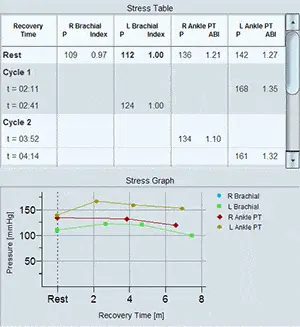
Figure 1 (Page 2) shows bilateral brachial, thigh, above knee, below knee and ankle (posterior tibial and dorsalis pedis) blood pressures, as well as common femoral, superficial femoral, popliteal, posterior tibial and dorsalis pedis artery Doppler waveforms.
Figure 2 (Page 3) shows bilateral thigh, above knee, below knee and ankle PVR. As Falcon Pro has 10 independent pressure channels, all PVR measurements can be made at the same time, hence completing all measurements in just a few seconds.
Figure 3 (Page 4) shows bilateral ankle PVR post exercise. Again, bilateral measurements for each ankle can be carried out at the same time.
Figure 4 (Page 5) shows bilateral ankle pressures and ABIs at rest and post treadmill exercise.

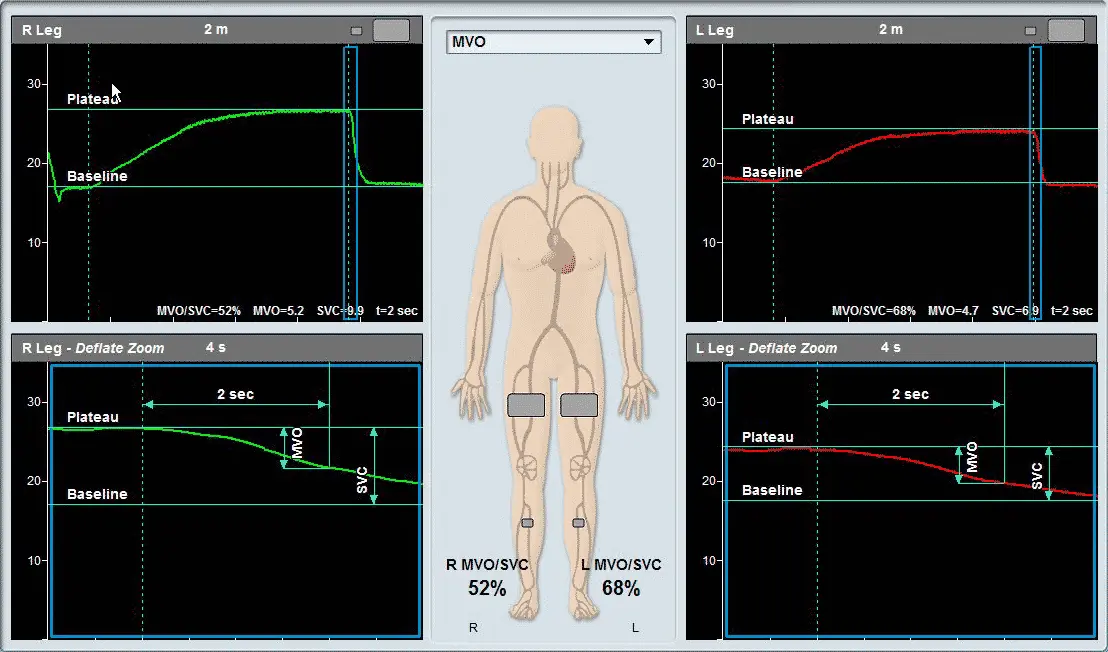
The Segmental Venous Capacitance / Maximum Venous Outflow (MVO/SVC) test is designed to assess lower limb venous outflow. The test is performed with the patient in supine position and the legs slightly elevated.
To perform the test, place a cuff on the thigh, and another cuff on the calf, record waveform obtained from cuff on the calf until a baseline is reached, rapidly inflate the thigh cuff to occlude veins until a plateau is reached, rapidly deflate the thigh cuff. Falcon Pro will automatically calculate and display MVO/SVC which is a rate calf cuff pressure decrease in pre-defined time frame of total pressure increase following thigh cuff inflation.
Figure is an example of bilateral MVO/SVC test. Both legs were measured simultaneously. The waveform above shows the calf cuff pressure changes during the test and the waveform below presents a zoom-in on the deflation part (blue rectangle). Decrease in MVO/SVC suggests venous obstruction.